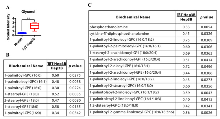
Figure 2.
Increased glycerol levels and decreased lysolipids and phospholipids in TβT-Hep3B cells. (A) Level of glycerol is depicted by a box plot with whiskers (min to max). Welch’s two-sample t-test was used to identify biochemicals that differed significantly between experimental groups (n = 5 for each group). ** p < 0.01. (B,C) Levels of lysolipids (B) and phospholipids (C) of TβT-Hep3 compared to Hep3B presented in fold. Welch’s two-sample t-test was used to identify biochemicals that differed significantly between experimental groups (n = 5 for each group, p-value indicated in the right column).

