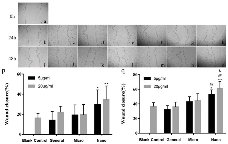
Figure 3.
Different concentration and particle sizes of pearl powder and their effects on keratinocyte migration: (a) injury at 0 h; (b,i) control group; (c,j) pearl powders with concentration of 5 μg/mL; (d,k) pearl powders with concentration of 20 μg/mL; (e,l) micron-sized pearl powders with concentration of 5 μg/mL; (f,m) micron-sized pearl powders with concentration of 20 μg/mL; (g,n) nano-sized pearl powders with concentration of 5 μg/mL; (h,o) nano-sized pearl powders with concentration of 20 μg/mL. Statistical data of injury closure percentage in wound scratch assay in (p) 1 day and (q) 2 days, determined by Image J software (U.S. National Institutes of Health, Bethesda, MD, USA, v1.51 23 April 2018). Reproduced from [4]. (* p < 0.05, vs. blank control; ** p < 0.01, vs. blank control; ## p < 0.01, vs. general; & p < 0.05, vs. micro).

