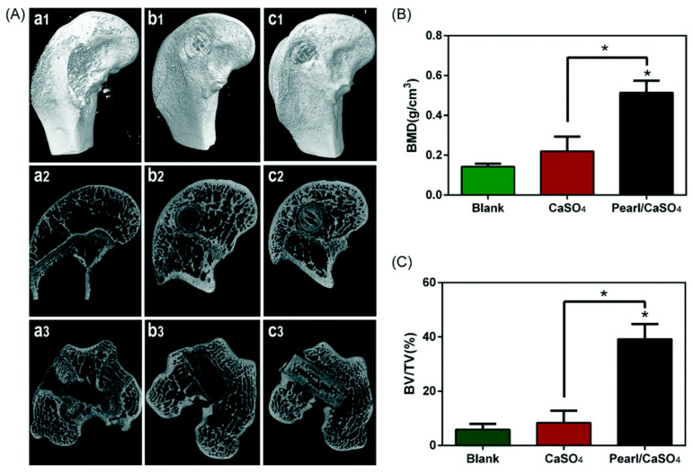
Figure 6.
Post-implantation evaluation of repaired skulls. (A) 3D images of (a1–a3) control, (b1–b3) scaffolds from CaSO4, and (c1–c3) scaffolds from pearl/CaSO4; morphometric analysis of (B) bone mineral density, and (C) bone volume/total volume. Reproduced from [79]. * p < 0.01.

