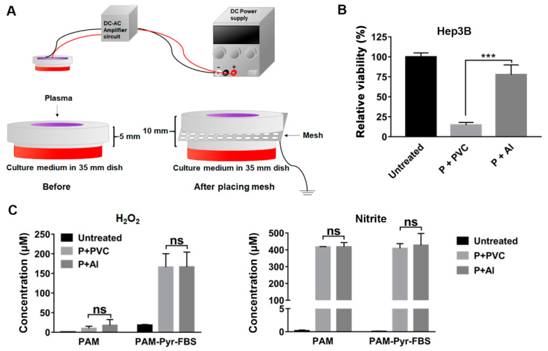
Figure 6.
Aluminum mesh, but not PVC mesh, significantly diminished the anti-proliferative effect of PAM. (A,B) The PAM was prepared by placing aluminum mesh (d, 1.5mm; t, 0.5 mm), PVC mesh (d, 1.2mm; t, 0.5 mm), or no mesh, respectively, and was treated with Hep3B cells; d, hole diameter; t, mesh thickness. (A) Schematic diagram of the preparation of PAM in the presence of mesh. (B) The viability of cells following each PAM treatment was measured after 72 h of the treatment using MTT assays. Relative viability was calculated as the ratio of the viability of treated to untreated cells observed at 72 h. (C) Concentrations of H2O2 and NO2− were measured in the PAM immediately after CAP exposure with PVC or Al metal mesh. The PAM containing both 1 mM sodium pyruvate (Pyr) and 10% FBS or without both 1 mM Pyr and 10% FBS was used. The concentrations of H2O2 were detected using Amplex® Red Hydrogen Peroxide assay and the concentrations of NO2− were detected using the Griess assay. Untreated culture medium was used as a control. (B,C) The results are plotted as mean ± SD of at least three independent experiments. *** p < 0.001 indicate significant difference; ns, not significant.

