
FIGURE 5.
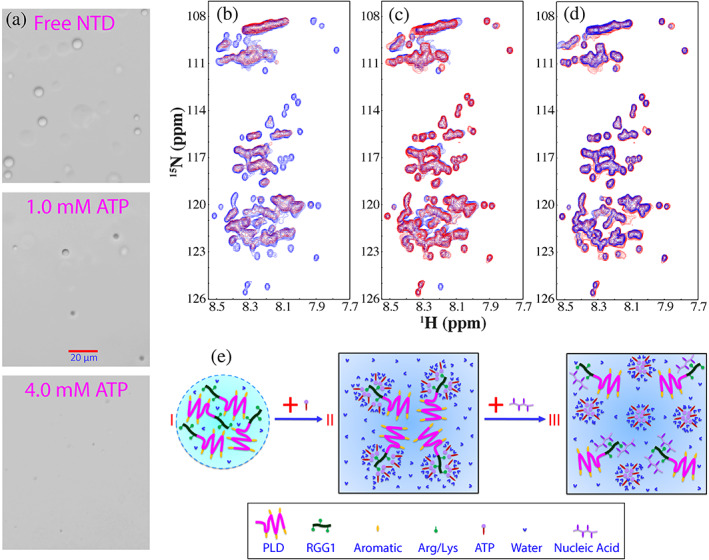
The modulation of LLPS of FUS NTD by ATP and ssDNA. (a) DIC images of dynamic droplets formed by FUS NTD in the presence of ATP at different concentrations. (b) HSQC spectra of FUS NTD in the absence (blue) and in the presence of ATP at 3 mM (red). (c) HSQC spectra of FUS NTD in the absence (blue), and in the presence of ATP at 3 mM with an extra addition of telomeric ssDNA (TssDNA) at 60 μM (red). (d) HSQC spectra of FUS NTD in the presence of TssDNA at a ratio of 1:5 (blue) and in the presence of both ATP at 3 mM and TssDNA at 60 μM (red). (e) A speculative model to illustrate: (1) FUS NTD undergoes LLPS resulting from the π‐cation or/and π‐π interactions between aromatic residues in PLD and Arg in RRG1; (2) the specific binding of ATP to Arg within RGG1 leads to dissolution of LLPS, which, however, can be displaced by ssDNA due to the much higher binding affinity of ssDNA