Figure 2.
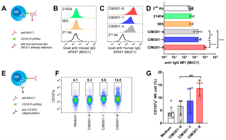
Humanized anti-MUC1 antibodies bind to CD16 on NK cells and induce degranulation. (A) Schematic overview outlining the detection of mAb binding via their Fc tail to the FcγRIIIa receptor (CD16) expressed on NK cells. (B) Binding of murine anti-MUC1 antibodies on primary human NK cells. Histograms show expression levels of the labeled anti-mouse IgG secondary antibodies used to detect MUC1 antibody binding. (C) Binding of humanized anti-MUC1 antibodies on primary NK cells. Experiment as in B, but using humanized anti-MUC1 antibodies, detected using anti-human IgG secondary antibodies. (D) Quantification of anti-MUC1 antibody binding to primary human NK cells. (E) Scheme depicting analysis of NK cell degranulation. (F) Flow cytometric analysis of NK cell degranulation following 4 h incubation of primary human NK cells with anti-MUC1 antibodies. One representative sample is shown with numbers indicating frequencies of cells within the indicated gate. (G) Quantification of the degranulation assay shown in (F). Bars in (D,G) show mean ± SD with individual data points as dots. Pooled data from seven independent experiments with different donors performed at different time points. Statistical analysis using one-way ANOVA plus Tukey’s multiple comparisons test. In (D), only biologically relevant comparisons were made. Not significant (n.s.); p < 0.01 (**); p < 0.0001 (****).

