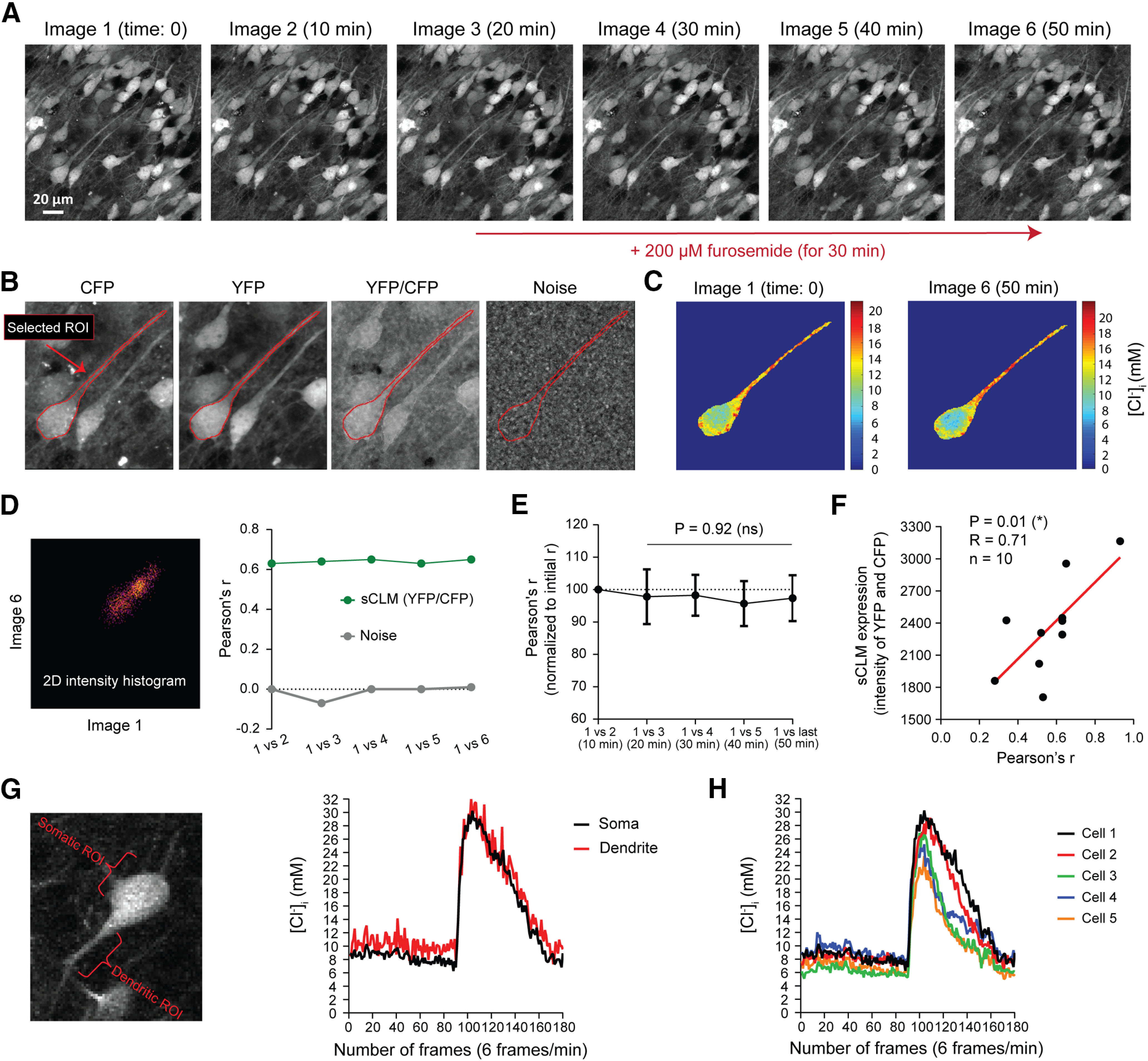
Figure 8.

Chloride microdomains are stable and are not defined by Cl– transport. A, Time-lapse images of a hippocampal pyramidal cell, expressing sCLM in the presence of 1 μm TTX before and after blocking KCC2 and NKCC1 with 200 μm furosemide. B, The stability of microdomains was measured by selecting an ROI and comparing YFP/CFP values in each pixel of that ROI over time. The same ROI was also imaged while the microscope shutter was closed to measure the noise level and pixel-by-pixel correlation of the ROI as a control. C, [Cl–]i in soma and dendrite of the selected ROI before and after 30 min application of furosemide. D, Pixel intensity correlation analysis in time-lapsed images was performed by generating 2D intensity histograms and calculating Pearson's r, using the Fiji plugin Coloc2. The colocalization correlation analysis of the [Cl–]i indicates that these dendritic Cl– microdomains remain stable before and after the application of furosemide. E, The change in spatial correlation of the sequential ratiometric images (normalized to the maximum Pearson's correlation value obtained at baseline) demonstrates that the spatial stability of Cl– microdomains does not change significantly over time after the application of 200 μm furosemide. F, The observed variability in Pearson's r in different experiments was significantly correlated with expression levels of sCLM. G, To demonstrate that small and large fluctuations in somatic and dendritic ROIs are detectable by sCLM imaging, sCLM-expressing cells were imaged before and after SLEs. G shows an example of a CA1 pyramidal neuron expressing sCLM, which was imaged for 30 min (6 frames/min; 180 total frames). Small changes in [Cl–]i can be detected at baseline (preictal period), as well as a large Cl– transient during an SLE in somatic and dendritic ROIs. H, Fluctuations in [Cl–]i in five different somatic ROIs before and during an SLE.
