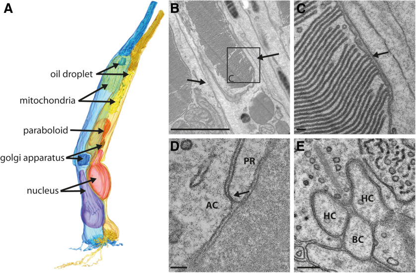
Figure 2.
Double-cone anatomy in the chicken retina. A, 3D reconstruction of both members of a double cone, the principal member (blue) and the accessory member (pale orange), from the outer segments containing densely packed disk membranes to the photoreceptor terminal where the signal transfer occurs. The oil droplets in both members (green) are located in the inner segments directly at the border to the outer segments. In the accessory member, multiple small oil droplets or granules could be found. The >200 mitochondria/cell (yellow) are densely packed in the ellipsoid part of the inner segment. The Golgi apparatus (red) is located in the myoid part of the inner segment close to the nucleus (magenta). The accessory member of the double cone additionally contains a paraboloid (dark orange) in the myoid part of the inner segment. B, Transmission electron microscopy (TEM) image of the outer segment of an accessory member from a double cone. Densely packed discs are visible as well as two of the multiple oil droplets and calyceal processes on both sides of the outer segment, as indicated by the arrows. C, Magnified area from B reveals the typical invaginations formed in the outer segments of cones (arrow). D, TEM images of the outer membranes of both double-cone members, which form junction-like structures (arrow) along the inner segments. E, TEM image of a ribbon synapse in the principal member of a double-cone terminal. HC, horizontal cell dendrite; BC, invaginating bipolar cell dendrite. Scale bars: B, 2 µm; C, D, 100 nm; E, 200 nm.

