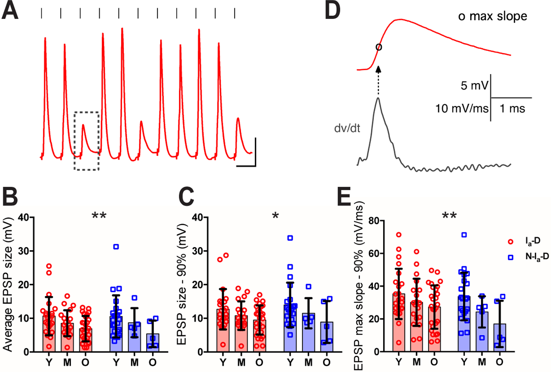
Figure 10. Evoked EPSPs decrease in amplitude with age during ARHL.

(A) Example response of a bushy neuron to show AN stimulation evoked spikes and EPSPs. Dashed rectangle: an example EPSP that failed to trigger any spike. Scale: 10 mV and 10 ms.
(B-C) Comparisons of bushy neurons among three age groups in average amplitude of EPSPs that failed to trigger spikes (B), and EPSP amplitude at 90th percentile (C).
(D) Maximum rising slope of the example EPSP from (A), calculated as the peak of the first derivative (dv/dt) during the rising phase.
(E) Comparison of EPSP maximum slope at 90th percentile.
Two-way ANOVA revealed significant age effect (Ia-D vs. Non-Ia-D) in B, C and E: *p < 0.05; **p < 0.01.
