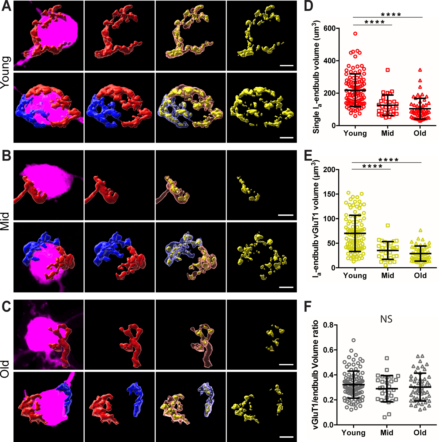
Figure 11. Synaptopathy of individual type Ia endbulb of Held synapses during ARHL.

(A) Representative morphology of individual type Ia endbulb of Held synapses in young mice. Panels from left to right: filled neurons (magenta) with reconstructed individual type Ia endbulbs, type Ia endbulbs alone, type Ia endbulbs (semi-transparent) with enclosed VGluT1-labled puncta (yellow), and VGluT1-labeled puncta alone. Top panels: example bushy neuron with only one type Ia endbulb of Held synapse. Bottom panels: example neuron with two type Ia endbulb of Held synapses, which are shown in red and blue respectively.
(B) Representative morphology of individual type Ia endbulb of Held synapses in middle-aged mice.
(C) Representative morphology of individual type Ia endbulb of Held synapses in old mice. Scales in A-C: 5 μm.
(D-F) Comparisons of individual type Ia endbulb volume (D), enclosed VGluT1-puncta volume (E), and VGluT1/endbulb volume ratio (F) among three age groups. Dunn’s multiple comparison test: NS, p > 0.05; ****p < 0.0001.
