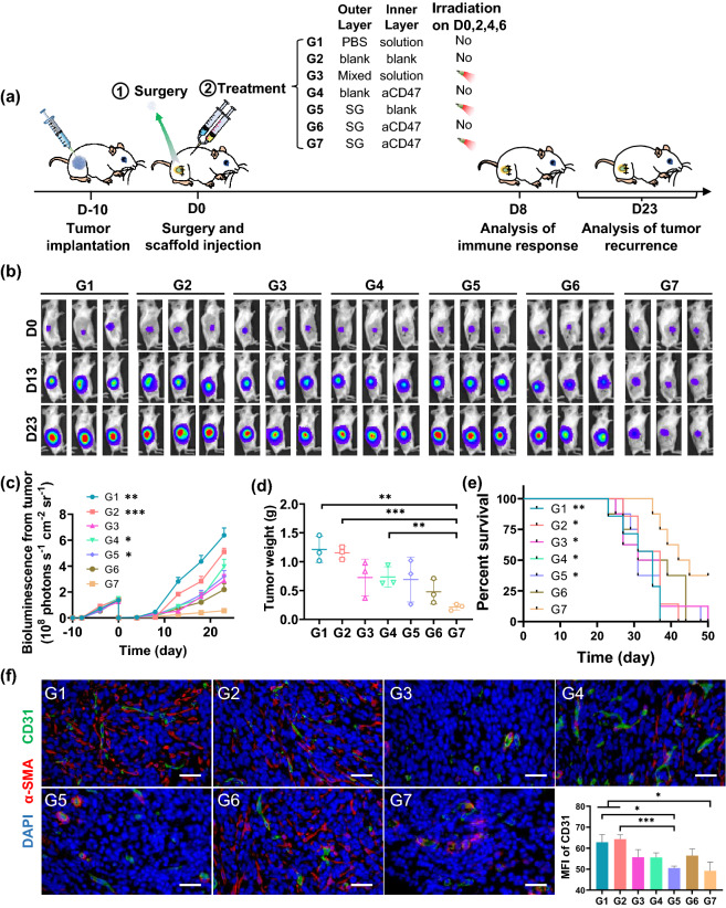
Fig. 4.
Sequential delivery of combined immunotherapy for inhibiting recurrence of 4T1 carcinoma tumor after surgery. a Schematic illustration of the animal experimental design. DLG outer layer: 50 μL; DLG inner layer: 50 μL; Dose of sorafenib: 2.5 mg kg−1; Dose of aCD47: 3.5 mg kg−1. b In vivo bioluminescence imaging of tumor resected mice receiving various treatments (G1–G7) after surgery. Three representative mice in each treatment group are shown. Images associated with Day 0 were taken on the day of surgery. c Tumor growth curves with the mean tumor volumes of 4T1 tumor-bearing BALB/c mice model. Data are shown as mean ± SEM (n = 5). Data difference is shown comparing to G7. d The recurrence tumor weights of different groups obtained from the 4T1-tumor resection mice on Day 23 after treatment. Data are shown as mean ± SEM (n = 3). e The survival percentages corresponding to the tumor volume of the 4T1-tumor resection mice (n = 8). Data difference is shown comparing to G7. f Immunofluorescence staining for tumor microvessels in tumor tissue on Day 8 after different treatments. The vascular endothelial cells were stained with the FITC-anti-CD31 antibody (green) and the vascular smooth muscle cells were stained with Cy5-anti-α-SMA antibody (red) and the cell nucleus was stained with DAPI (blue) (Scale bar: 100 μm). The comparison of two groups was followed by student’s t test (two-tailed). *p < 0.05, **p < 0.01, and ***p < 0.001

