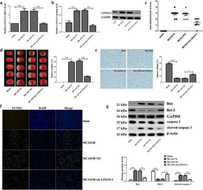
Fig. 2.
Effect of sh-LINGO-1 on I/R-induced MACO mice model. a, b LINGO-1 mRNA and protein expression were detected by qRT-PCR and WB at 7 days after MCAO. c Neurobehavioral score determination (Longa scoring system) Sham, MCAO/R (7 days), MCAO/R + NC, MCAO/R + sh-LINGO-1 (lentiviral vectors) groups. d Brain infarct region in TTC staining. Red: Non-ischemic area; white: ischemic area. Percentage of infarct volume after TTC staining. e Nissl staining. f TUNEL staining. g Expression of apoptosis-related proteins Bax, Bcl-2, Caspase 3, and Cleaved caspase 3 were detected by WB. Data are expressed as mean ± SEM and *p < 0.05; **p < 0.01; ***p < 0.001

