Figure 1.

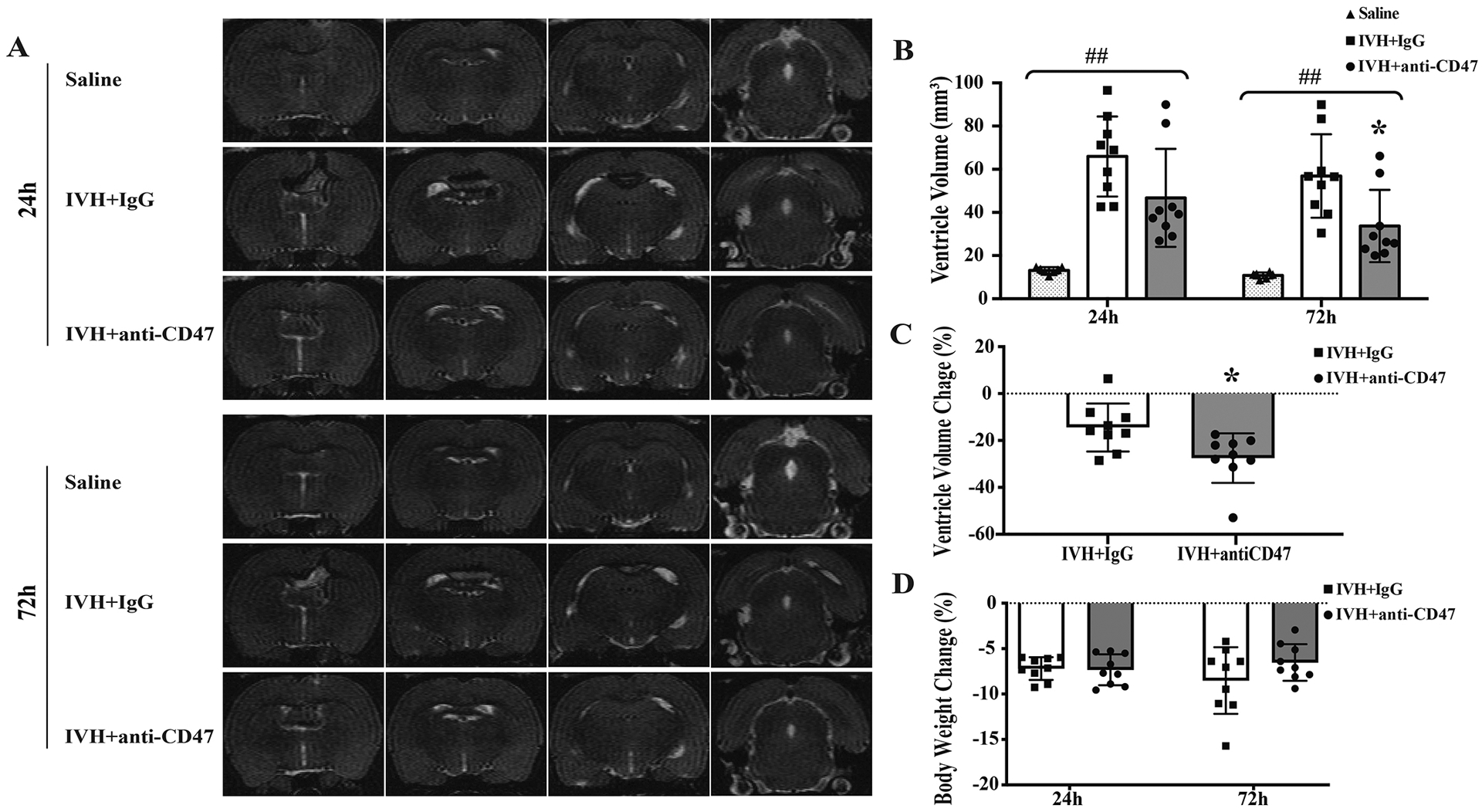
(A) Representative T2-weighted MRI scans at 24 and 72 hours after intraventricular injection of 200 μl saline (Saline) as control or 200 μl autologous blood with IgG (IVH+IgG) or CD47 blocking antibody (IVH+anti-CD47) in F344 rats. Note the significant ventricular dilation in rats with IVH compared to saline control. (B) Ventricular volumes were quantified using the T2-weighted images in the three groups. The CD47 blocking antibody significantly reduced the ventricular dilation after IVH. (C) The percent changes in ventricular dilation between 24 and 72 hours also significantly increased the CD47 antibody group. (D) Percentage changes from pre-IVH body weights 24 and 72 hours after IVH. For all graphs, data are shown as mean ± SD; n = 6–7 in saline group, n = 9 in IVH groups; ## p< 0.01 between groups by one-way ANOVA with Tukey post hoc test, * p < 0.05 IVH+anti-CD47 vs. IVH+IgG group.
