Figure 7.

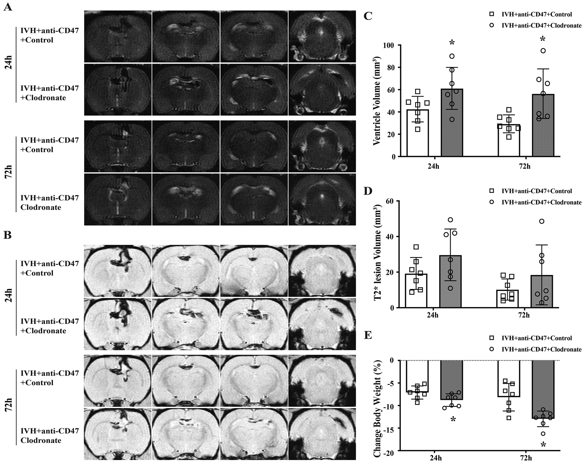
(A) Representative T2-weighted MRI scans at 24 and 72 hours after intraventricular injection of 200 μl autologous blood with CD47 blocking antibody plus clodronate liposomes (IVH+anti-CD47+Clodronate) or control liposomes (IVH+anti-CD47+Control) in F344 rats. (B) Representative images of T2*-weighted MRI scans at 24 and 72 hours after intraventricular injection of 200 μl autologous blood with CD47 blocking antibody plus clodronate liposomes (IVH+anti-CD47+Clodronate) or control liposomes (IVH+anti-CD47+Control) in F344 rats. (C) Ventricular volumes were quantified using the T2-weighted images. The clodronate liposomes treatment significantly increased ventricular dilation after IVH. (D) Intraventricular lesion volumes were quantified using T2*-weighted images at 24- and 72-hours post-surgery. (E) Weight changes from pre-IVH to 24 and 72 hours after IVH. For all graphs, data are shown as mean ± SD; n = 7 per group; * p < 0.05 IVH+anti-CD47+Clodronate vs. IVH+anti-CD47+Control group.
