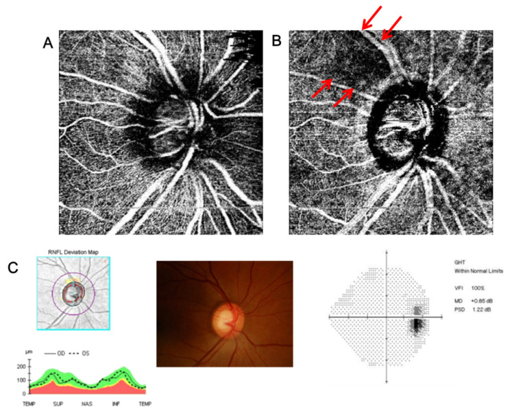
Figure 1.
(A) Superficial peripapillary OCT angiography in the superficial layer (from the retinal nerve fiber layer (RNFL) to 130 μm below the internal limiting membrane (ILM) including the ganglion cell-inner plexiform (GCIPL) layer). (B) Deep peripapillary OCT angiography in the deep layer (from 130 μm below the ILM to the basement membrane including the inner nuclear layer (INL)). (C) RNFL OCT and visual field test with increased cup-disc ratio (CDR) which indicate glaucoma suspect. There are subjects among glaucoma suspect patients with wedge-shaped VD defect in the deep layer of peripapillary area (indicated as arrows in B), even though the vessel density in the superficial layer is unaffected (intact vascular status in A).

