Figure 3.
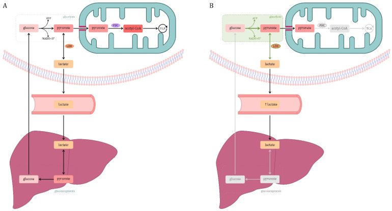
Glucose metabolism. (A) Physiological conditions. Glycolysis is the conversion of glucose into pyruvate. In some cases, for example, when glycolysis occurs anaerobically or in cells incapable of pyruvate oxidation, pyruvate may be converted to lactate by the enzyme lactate dehydrogenase (LDH). Blood lactate (mainly formed in skeletal muscle) is taken up by the liver, converted to pyruvate and incorporated into the gluconeogenesis pathway (Cori cycle). In other cases, the pyruvate formed in the cytosol is transported to the mitochondria by a proton symporter. In the mitochondrion, pyruvate undergoes oxidative decarboxylation to acetyl-CoA mediated by the multi-enzyme pyruvate dehydrogenase complex (PDC). Oxidation of pyruvate to acetyl-CoA connects glycolysis with the tricarboxylic acid (TCA) cycle [74]. (B) Sepsis. This is due, inter alia, to a decrease in pyruvate dehydrogenase complex (PDC) activity. One explanation is the phosphorylation of the enzyme by pyruvate dehydrogenase kinases (PDKs) and its decreased expression in sepsis. Moreover, in sepsis, the hypoxia-inducible factor 1α (HIF-1α) is activated, which is one of the main mediators of glycolysis. HIF-1α regulates the expression of genes of enzymes related to glycolysis (hexokinase, phosphofructokinase-1, glucose-6-phosphate dehydrogenase, lactate dehydrogenase, pyruvate dehydrogenase kinase, glutamate transporter-1). The intensification of glycolysis in combination with the failure of entering pyruvate to the tricarboxylic acid (TCA) cycle increases the formation of lactate. A decrease in hepatic lactate-based gluconeogenesis has been reported. This increases the production of lactate, while blocking the main routes of its disposal, which results in an increase in its concentration in the blood. Legend: gray color—inefficient cellular processes; green color—increasing the activity of the process or enzyme.

