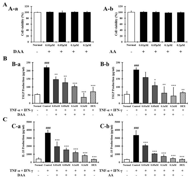
Figure 2.
Effect of DAA and AA on HaCaT cell viability and the levels of AD-related cytokines and chemokines in HaCaT cells. (A) The viability of HaCaT cells treated with DAA and AA. (B) DAA and AA inhibited the secretion of TSLP by HaCaT cells treated with both TNF-α and IFN-γ. (C) DAA and AA inhibited the secretion of IL-33 by HaCaT cells treated with both TNF-α and IFN-γ. The production of cytokines and chemokines in the supernatant of HaCaT cells was analyzed by ELISA. The results are expressed as the mean ± SD (n = 4). ### p < 0.001 vs. normal; * p < 0.05, ** p < 0.01 and *** p < 0.001 vs. control; DAA was used at 0.01 µM, 0.05 µM, 0.1 µM, and 0.2 µM. DEX, the positive control, was used at 0.1 µM.

