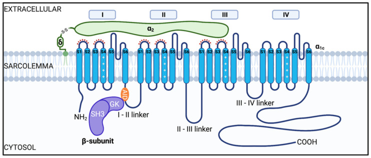
Figure 1.
Illustration of the cardiac voltage gated L-type CaV1.2 channel complex. The channel consists of a pore-forming CaVα1C subunit and auxiliary subunits CaVβ and CaVα2δ. The CaVα1c is composed of four homologous repeat domains (I-IV), each having six transmembrane spanning segments (S1–S6, shown in blue). S1–S4 comprise the voltage sensing domain (VSD) and S5–S6 form the pore domain (PD). CaVβ subunits (depicted in purple) are composed of a SH3, HOOK, and GK domain. Interaction of CaVβ with the CaVα1c occurs between the GK domain on CaVβ subunits and the alpha interaction domain (AID) on the I-II linker (orange). CaVα2δ subunits are proposed to interact with the extracellular loops of domains I-III as highlighted by the red dashed lines [10,11,12,13].

