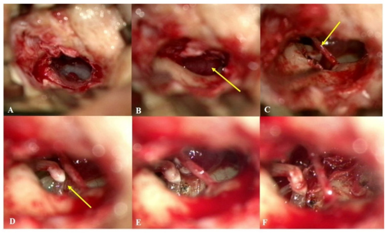
Figure 3.
Intraoperative microscopic images of subject 8. (A) An endaural approach was chosen in this case. (B) After the elevation of a tympanomeatal flap, a round, reddish tumor (arrow) was observed in the right mesotympanum. (C) The chorda tympani nerve (arrow) was mobilized to improve the exposure of the mass. (D) The incudostapedial (I-S) joint (arrow) was located and separated to protect the stapes while mobilizing the mass abutting the incus. (E) A 6 mm-sized glomus tympanicum tumor with a stalk arising from the promontory was observed. (F) After the feeding vessel was cauterized with a CO2 laser, the tumor was fully removed en-bloc without any injuries to critical structures or severe hemorrhage. The I-S joint was immediately reconnected using bone cement.

