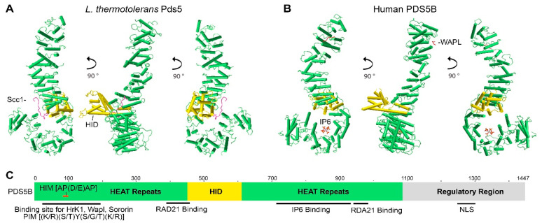
Figure 1.
Structure and characteristics of the PDS5 protein. Crystal structure images of (A) L. thermotolerans Pds5 (PDB ID: 5F0O) and (B) human PDS5B (PDB ID: 5HDT) were generated by using NCBI’s web-based 3D structure viewer iCn3D. PDS5 is a hook-like molecule consisting of HEAT repeats (green sticks) with helical insert domain (HID, golden sticks) extruding on one side of the hook. Binding of Scc1/Rad21 (pink line) to L. thermotolerans Pds5 is shown in (A). Binding of WAPL (red line) to the C-terminus of human PDS5B and IP6 to the bottom of PDS5B hook-like structure are shown in (B). (C) Schematic drawing shows PDS5 molecular features. The relative site is based on human PDS5B. Hrk1 interacting motif (HIM) on PDS5 N-terminus interacts with the PDS5 interacting motif (PIM) on HrK1, WAPL, and sororin. RAD21 and IP6 interact with PDS5 in the middle region. Nuclear localization signal (NLS) is on the C-termini of PDS5A and PDS5B (Refer to Figure 2).

