Figure 1.
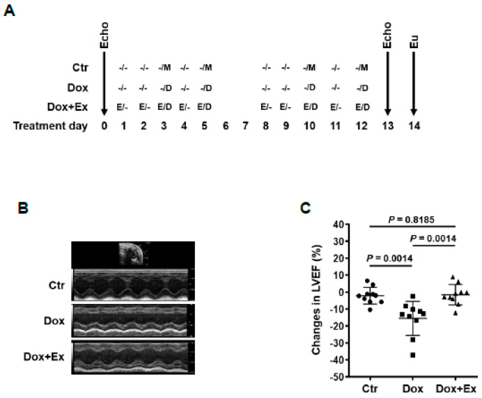
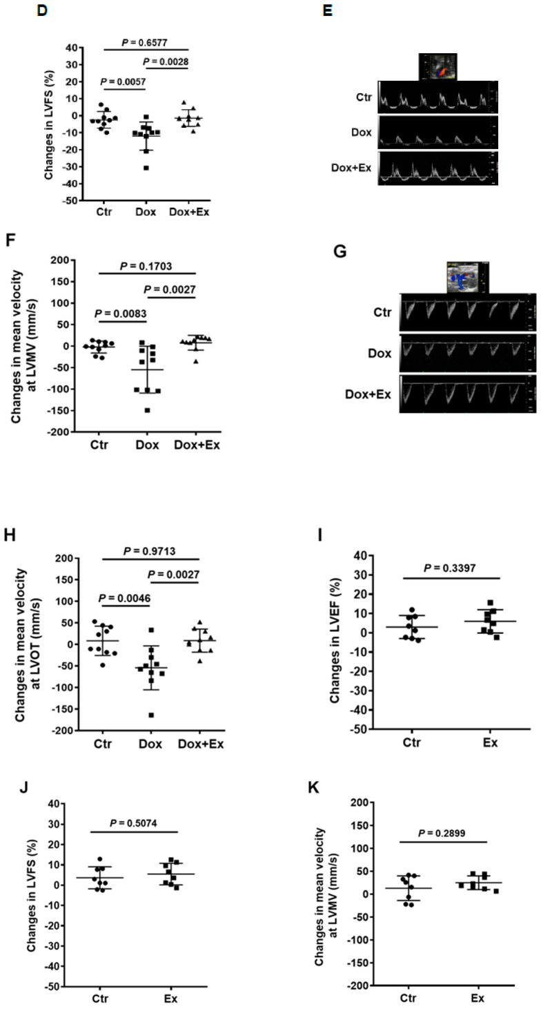
Exercise prevents Dox-induced decreases in cardiac function and blood flow. (A) Schematic representation of experiment design and mouse treatment. (B,E,G) Representative Echo images of M-mode of parasternal short axis (PSAX) view of hearts (B), PW Doppler waveform of trans-mitral blood flow in hearts (E), and PW Doppler waveform of transverse aorta blood flow in hearts (G) from Ctr, Dox, and Dox+Ex group mice. (C,D,F,H) Echo data show changes in LVEF (C), LVFS (D), mean velocity at LVMV (F), and mean velocity at LVOT (H) after treatment in Ctr, Dox, and Dox+Ex group mice. N = 10 mice/group. P values are indicated by the GraphPad t test. Ctr vs Dox+Ex, not significant statistically. (I–K) Echo data show changes in LVEF (I), LVFS (J), and mean velocity at LVMV (K) after treatment in Ctr and Ex group mice. N = 8 mice/group. P values are indicated by the GraphPad t test. Ctr vs Ex, not significant statistically. LVEF, left ventricular ejection fraction; LVFS, left ventricular fractional shortening; LVMV, left ventricular mitral valve; LVOT, left ventricular outflow tract in the ascending aorta; PW Doppler, pulse wave Doppler; Ctr, control; M, mock; Ex/E, exercise; Dox/D, doxorubicin; Echo, echocardiography; Eu, euthanize.


