Figure 3.
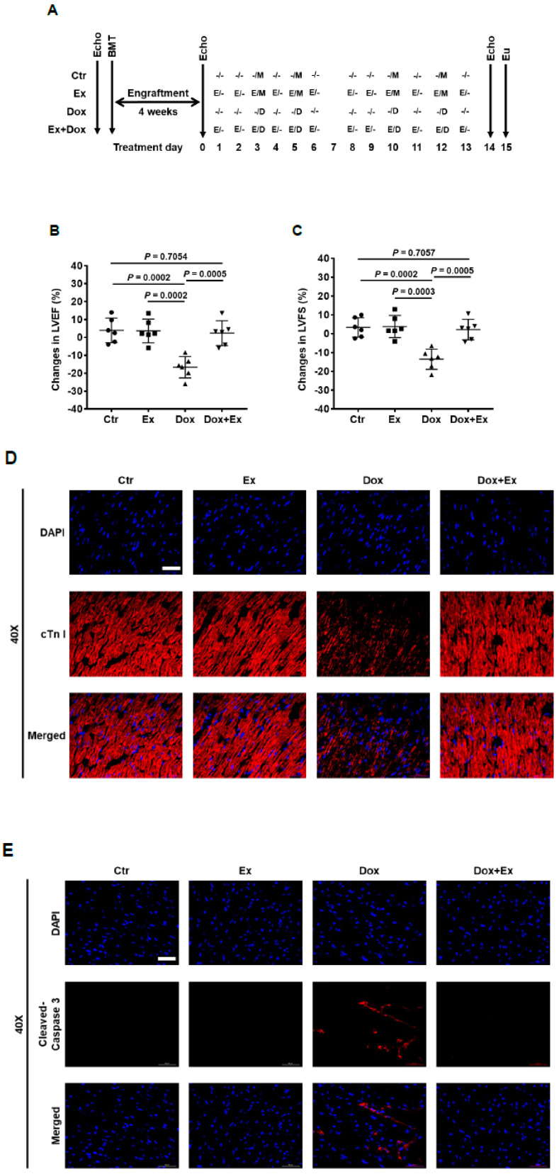
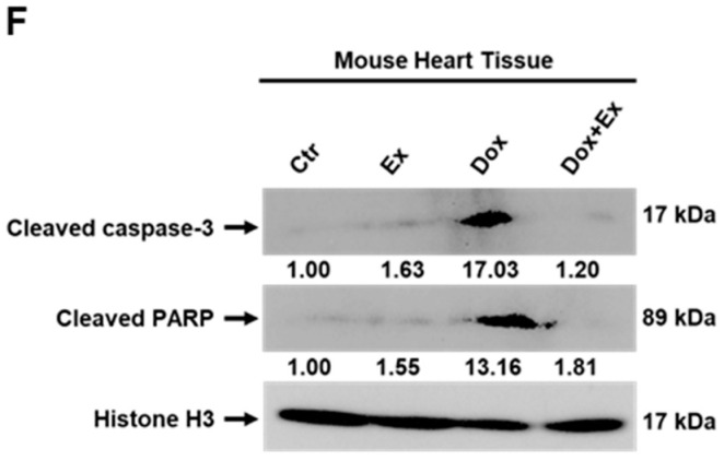
Exercise inhibits Dox-induced cardiomyocyte apoptosis. (A) Schematic representation of experiment design of BMT and mouse treatment. (B,C) Echo data show changes in LVEF and LVFS before and after treatment in Ctr, Ex, Dox, and Dox+Ex group mice. N = 6 mice/group. P values are indicated by the GraphPad t test. Ctr vs Dox+Ex, not statistically significant. (D) Representative immunofluorescence images of heart sections stained for DAPI (blue) and cTn I (red) from Ctr, Ex, Dox, and Dox+Ex group mice. Magnification, 40×; Scale bar, 50 µm. (E) Representative immunofluorescence images of heart sections stained for DAPI (blue) and cleaved caspase-3 (red) from Ctr, Ex, Dox, and Dox+Ex group mice. Magnification, 40×; Scale bar, 50 µm. (F) Western blot analysis for cleaved caspase-3, cleaved PARP, and histone H3 expression in hearts from Ctr, Ex, Dox, and Dox+Ex group mice. LVEF, left ventricular ejection fraction; LVFS, left ventricular fractional shortening; Ctr, control; M, mock; Ex/E, exercise; Dox/D, doxorubicin; Echo, echocardiography; Eu, euthanize; BMT, bone marrow transplant.


