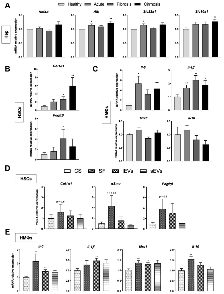
Figure 4.
Effects of LSECs secretome on neighbouring liver cells during CLD. mRNA expression of specific phenotype markers in hepatocytes (A), HSCs (B) and HMΦs (C) incubated for 24 h with the complete secretome (CS) of LSECs isolated at each stage of CLD. mRNA expression of phenotype markers in HSCs (D) and HMΦs (E) after 24 h of incubation with the different sub-fractions of cirrhotic LSECs secretome: large extracellular vesicles (lEVs), small EVs (sEVs) and soluble factors (SF), normalized to CS-receiving cells. Results derive from n = 3–5 independent experiments. * p < 0.05 and ** p < 0.01 on t test (Mann-Whitney U test for non-parametric variables) for differences between each group and the CS group.

