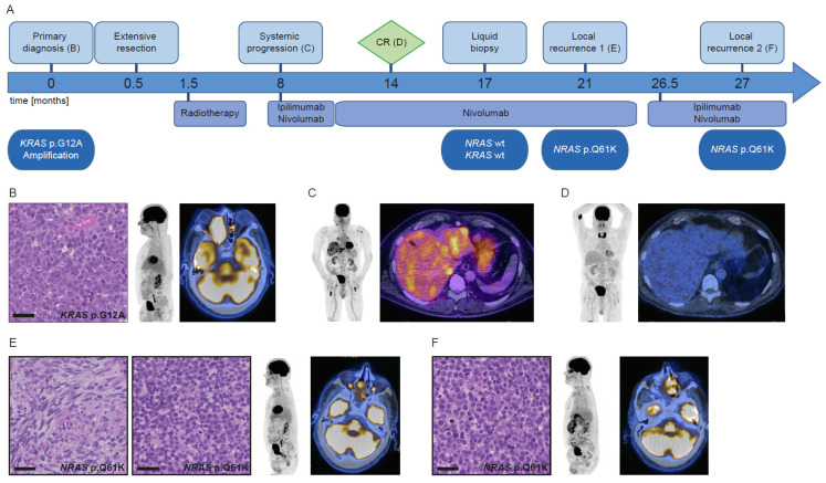
Figure 2.
Course of disease, treatment, and morpho-molecular workup of patient 2. (A) Timeline. (B) Histology of the primary tumor; PET MIP display and fused PET/CT image at primary diagnosis. (C) PET MIP display and fused PET/CT image at the start of immunotherapy. (D) PET MIP display and fused PET/CT image at CR. (E) Histology of the first local recurrence, left: spindle cell shaped morphology, right: monomorphic epithelioid morphology; PET MIP display and fused PET/CT image at first local recurrence. (F) Histology of the second local recurrence; PET MIP display and fused PET/CT image at second local recurrence. Scale bar: 40 µm. wt: wild type, CR: complete remission.

