Figure 1.
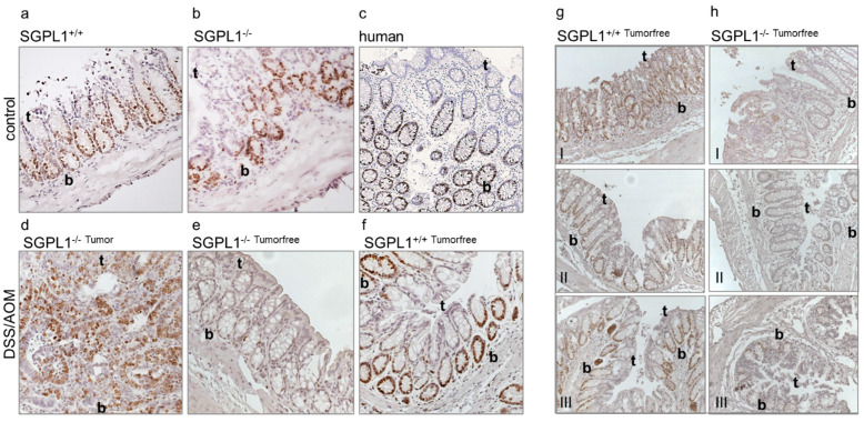
Anti-Ki-67 staining of (a) murine colon epithelium (n = 5), (b) murine SGPL1 knockout colon epithelium (n = 5), (c) human colon epithelium (n = 4), (d) murine SGPL1 knockout colon tumor after CAC induction (9 weeks) (n = 5), (e) murine SGPL1 KO colon epithelium (n = 5), (f) murine colon epithelium after CAC induction (9 weeks) (n = 5 (magnification 40×). Anti-Ki-67 staining (n = 3) of (g) wildtype and (h) SGPL1 knockout colon sections in different locations (I distal colon, II mid colon, III proximal colon); (magnification 20×); CAC = colitis associated colon cancer; in image: t = Crypt tip; b = crypt bottom.

