Figure 6.
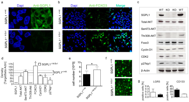
Anti-FOXO3-staining (n = 3) of (a) SGPL1+/+M. (magnification 60×) and (b) stable SGPL1-Exon1 knockout DLD-1 cells (SGPL1−/−M.Ex1); (magnification 40×) (c) Western blot analysis (n = 3), (d) protein quantification, (e) cell expansion within 72 h (n = 8), and (f) Anti-Ki-67 staining of SGPL1+/+ and SGPL1−/−M.Ex1 (n = 3); (magnification 40×); (g) FACS analysis of LGR5 and CD133 positive cells (n = 2). Significances: * p ≤ 0.05; *** p ≤ 0.001.

