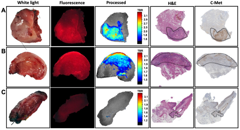
Figure 3.
The correlation between c-Met targeted fluorescence and immunohistochemistry. Overview of imaging and pathological assessment of three different samples showing ex vivo white light, Cy5-based fluorescence imaging of the incubated tissue (fluorescence in red), real-time image processing of the fluorescence signal (including video representation of tumor-to-background ratio (TBR)), H&E staining and c-MET immunohistochemistry. (A) superficial tumor localization, (B) tumor presence on the cleavage plane and (C) a sample without tumor presence on the cleavage plane. Delineation of the tumor (dashed) was based on H&E.

