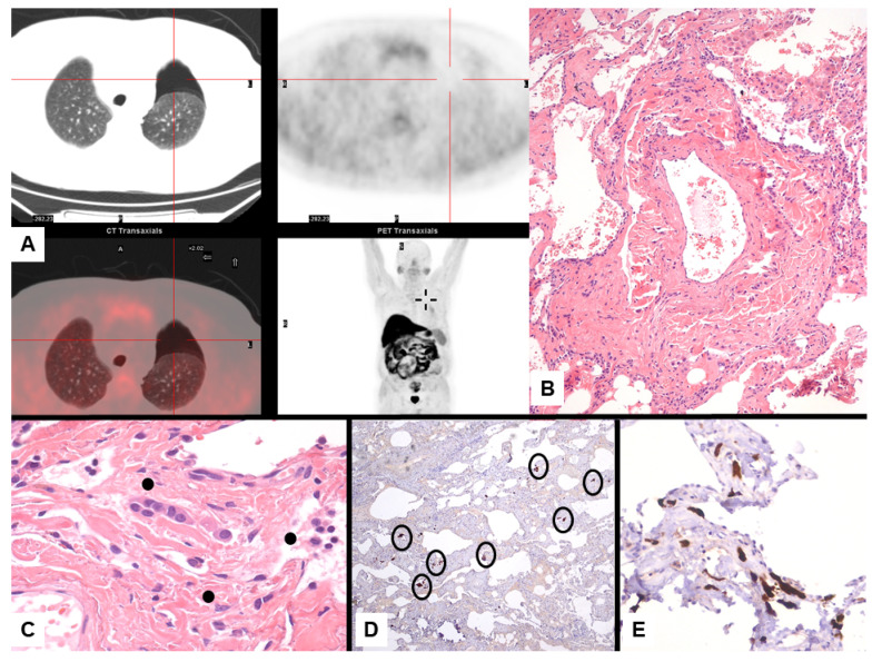
Figure 3.
Mesothelioma presenting with left pneumothorax at imaging study with PET/CT scan without evidence of pleural thickening (A). Lung resection of the left apex shows interstitial fibrosis with tiny, scattered, bland-looking proliferation of epithelioid cells (B) partially lining alveoli and merging in the fibrosis (C, dots). Calretinin staining highlights the mesothelial differentiation of epithelioid cell nests randomly scattered through the lung parenchyma (D, circles) lining the alveolar spaces (E).

