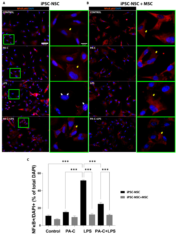
Figure 4.
PA-C pre-treatment and MSC co-culture prevent NF-κB translocation/activation in iPSC-NSC following pro-inflammatory LPS insult. (A,B). Representative immunostaining images of the NF-κB inflammatory marker (orange) and nuclear staining DAPI (blue) in iPSC-NSC monoculture (iPSC-NSC) (A) and MSC co-culture (iPSC-NSC + MSC) (B) incubated for 24 h with 10 µM PA-C or vehicle and then incubated with LPS (1 µg/mL) or vehicle for 24 h (scale bar = 100 µm). Green squares show higher magnification of the nuclei (scale bar = 20 µm), with white and yellow arrows indicating nuclear and cytosolic NF-κB, respectively. (C). Quantification of NF-κB translocation by quantifying iPSC-NSC with nuclear NF-κB (NF-κB+/DAPI+) expressed as a percentage of the total number of iPSC-NSC by DAPI. Data expressed as mean ± S.E.M. (at least eight random fields from three independent experiments) determined by one-way ANOVA with Tukey’s multiple comparison test (*** p < 0.001 as indicated).

