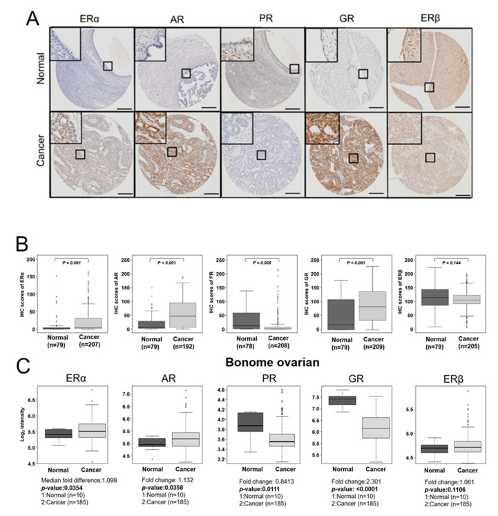
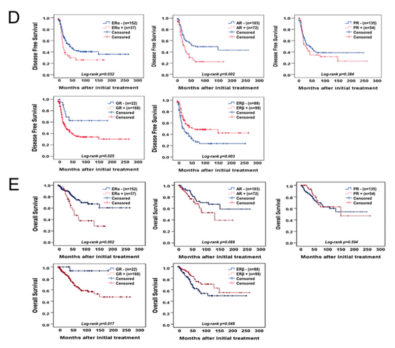
Figure 1.
Expression of hormone receptors in EOC tissues. (A) Representative immunohistochemical staining images of ERα, AR, PR, GR, and ERβ in nonadjacent ovarian epithelial tissues (Normal) and epithelial ovarian cancer (Cancer) tissue samples (scale bar: 50 μm). (B) Boxplots of IHC staining data (histoscores) comparing between nonadjacent ovarian epithelial tissues (Normal) and epithelial ovarian cancer (Cancer) tissue samples of each hormone receptor subtype. Histoscores were calculated based on staining intensity and the area of positive staining (C) Publicly available data on the mRNA expression of ERα, AR, PR, GR, and ERβ were obtained from the GEO data (GSE26712). A Mann–Whitney U-test or Kruskal–Wallis test was used to compare the mRNA expression level of each hormone receptor. (D) The DFS of patients with EOC depends on ERα, AR, PR, GR, and ERβ expression. For the DFS analysis, 189 patients with EOC for ERα, 175 patients with EOC for AR, 189 patients with EOC for PR, 190 patients with EOC for GR, and 187 patients with EOC for ERβ were included. (E) The OS of patients with EOC depends on ERα, AR, PR, GR, and ERβ expression. For the OS analysis, 189 patients with EOC for ERα, 175 patients with EOC for AR, 189 patients with EOC for PR, 190 patients with EOC for GR, and 187 patients with EOC for ERβ were included. The cut-off value of ERα was over 49.2 of the IHC score, cut-off value of AR was over 10.85 of the IHC score, cut-off value of PR was over 21.18 of the IHC score, and the cut-off value of GR was over 8.65 of the IHC score.


