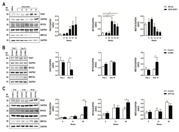
Figure 3.
Altered expression of PAX7, MYOG, and MEF2A proteins in G93A-SOD1, Δ7SMA, and AR113Q mouse muscle as disease progresses. Representative western blot analysis of PAX7, MYOG, and MEF2A proteins in gastrocnemius muscle tissue of (A) G93A-SOD1 (black bars), (B) Δ7SMA (black bars), and (C) AR113Q (black bars) mice and control mice (white bars) (n = 3 mice per group) with relative densitometric analysis. Density values are reported as mean ± SEM, corrected for background and normalized to GAPDH control. * p < 0.05, Mann-Whitney test.

