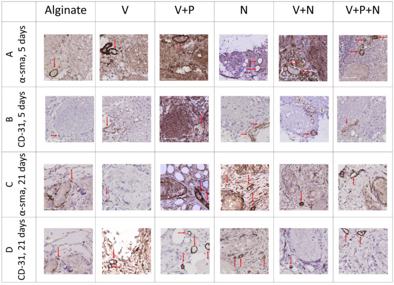
Figure 4.
Impact of NP supplementation on the vascularization of mice testicular tissue auto-transplanted for 5 and 21 days. Mouse testicular tissue fragments were encapsulated in alginate supplemented with five different combinations in addition to a negative control (Alginate, V, V+P, N, V+N and V+P+N) and the grafts were stained after 5 and 21 days for α-SMA and CD31. Red arrows highlight positive vascular structures. Scale bar = 60 μm. n = 3.

