Figure 2.
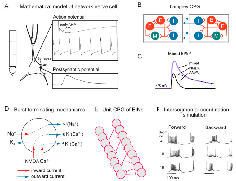
Cellular properties explored in simulations of the lamprey spinal locomotor networks. (A) The morphology of the CPG neurons is captured using a five-compartment model consisting of an initial segment, a soma and a dendritic tree. Active ion currents are modeled using a Hodgkin–Huxley formalism. Both ion channels involved in spiking behavior (Na+, K+) and slower Ca2+ or potassium-dependent processes (KCa, KNa) are modeled based on available data. Spiking behavior of a CPG neuron. Spike frequency can be regulated by the AHP (afterhyperpolarization). (B) The segmental locomotor network in lamprey. Red, excitatory interacting interneurons excite motoneurons (green) and commissural inhibitory interneurons (blue) that inhibit all neurons on the contralateral side. (C) Fast synaptic transmission is included in the form of excitatory glutamatergic (AMPA and voltage-dependent NMDA) and inhibitory glycinergic inputs. (D) Main ionic membrane and synaptic currents considered to be important during activation within the CPG network. Slower processes can cause spike frequency adaptation, such as Ca2+ accumulation during ongoing spiking and resulting activation of KCa. (E) Unilateral CPG activity can be evoked in local networks of excitatory interneurons (EINs). This basic EIN network can sustain the rhythm seen in ventral roots in vitro during evoked locomotor activity. The left–right alternating activity requires the presence of contralateral inhibition provided by glycinergic interneurons (see below). (F) The burst activity along the simulated spinal cord is illustrated in segments 4, 10 and 16. Note the sequential activation of the different segments.

