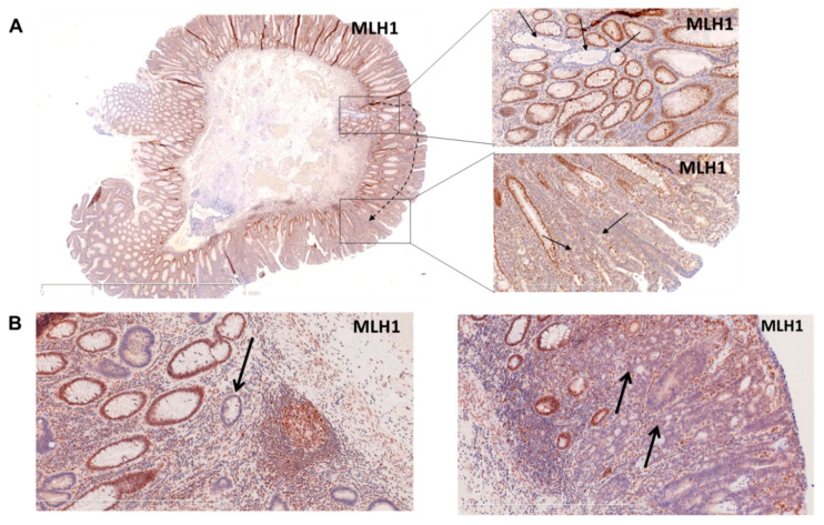
Figure 2.
Histology images of tumor specimens with MMR-deficient crypt foci. (A) Resection sample with carcinoma in situ arising presumably from an MMR-deficient crypt. On the left panel, the overview of the resected sample (MLH1 staining); on the right upper panel, higher magnification of the MMR-deficient crypt (MLH1 staining); on the right lower panel, higher magnification of carcinoma in situ (MLH1 staining). (B) MLH1 staining revealing an MMR-deficient crypt (indicated by an arrow), on the left and another region of the same sample showing a non-invasive carcinoma in situ (indicated by arrows) on the right panel.

