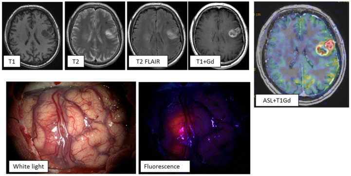
Figure 3.
MRI study of a patient with multiple primary glioblastomas. ASL perfusion in the tumor of the left frontal lobe revealed areas of hyperperfusion: TBF = 289.7 mL/100 g/min. Tumor demonstrated vivid contrast enhancement. During surgery using fluorescence diagnostics, this tumor showed an intense glow. Gd—gadolinium.

