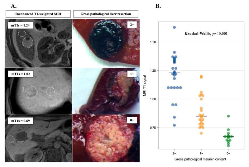
Figure 1.
Association between gross pathological and MRI melanin quantification. (A) Illustrative examples showing the correlation between preoperative, unenhanced, T1-weighted MRI and postoperative gross pathological scoring of pigmentation in uveal melanoma liver metastases. White dashed lines indicate regions of interest that were manually drawn in tumors and in adjacent liver parenchyma. Melanin pathological quantification was assessed visually and graded as follows: 0+ (hypopigmented), 1+ (mixed pigmented), and 2+ (strongly pigmented). (B) Correlation of gross pathological features and MRI melanin quantification. Horizontal lines indicate median values.

