Figure 6.
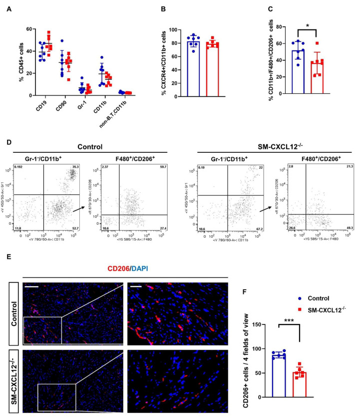
Cardiac M2-like macrophages were declined in SM-CXCL12−/− mice. (A) Bar graph showing flow cytometric analysis of CD45+ leukocyte subsets gated and quantified for CD19+ B lymphocytes, CD90+ T lymphocytes, Gr-1 granulocytes, CD11b monocytes/macrophages, n = 7. (B) Bar graph showing the percentage of CXCR4 + /CD11b+ cells in the hearts of control and cKO mice, n = 7. (C) Quantification of cardiac Gr-1-/CD11b+/F480+/CD206+ cells showing a significant reduction in SM-CXCL12−/− mice, n = 7. * p < 0.05 from Student’s t-test. (D) Representative gating strategy and scatter plots used to identify M2-like macrophage populations (Gr-1-/CD11b+/F480+/CD206+): For quantification of M2 macrophages, granulocyte negative CD11b+ cells were gated and stained for F480 and CD206. (E) Immunofluorescence labeling against CD206/DAPI confirmed a significant reduction of CD206+ cells in the myocardium of cKO mice. (F) Bar graph representing the quantification of cardiac CD206+ M2 like cells in the hearts of control and mutant mice, n = 6. Scale bar, 200 µm. *** p < 0.001 from Student’s t-test. Data represent mean ± SD.

