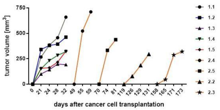
Figure 4.
After tumor cell injection of MDA-MB-231 (mouse #1.1 to #1.5) and MDA-hyb5 (mouse #2.2 to #2.5) into the NODscid mice, the tumors first became detectable at the indicated time points. Continuous tumor growth and detection of appropriate sizes were measured using a digital caliper. Progressive tumor volumes relative to the corresponding time points were calculated with the longitudinal diameter (length) and the transverse diameter (width) in the modified ellipsoidal formula: volume = π/6 × width × (length)2, as previously reported [38,39].

