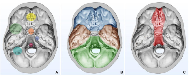
Figure 2.
(A) Relative frequencies in SBM locations are stratified and shown with circles of progressively wider diameter; (B) Topological subdivision of skull base: anterior skull base is shown in blue, middle skull base is shown in brown, and posterior skull base is shown in green; (C) Anatomical area for which endoscopic endonasal approach can be used.

