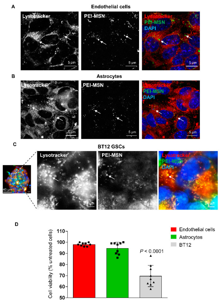
Figure 9.
The PEI-MSNs cross the BBTB in vitro. (A–C) Confocal images of the colocalization of PEI-MSN (green) with the Lysotracker dye (red) in the endothelial (A), astrocytic (B), and GSC (C) compartments 24 h after addition of the PEI-MSN. Images in (C) are magnified from a BT-12 gliosphere (squared region of interest on the left panel). (D) Cell viability measured by MTT after 72 h. Values are normalized to the control conditions (cells isolated from untreated BBTB). n = 3, three pooled experiments. p-value calculated with one-way ANOVA with Sidak’s multiple comparison test.

