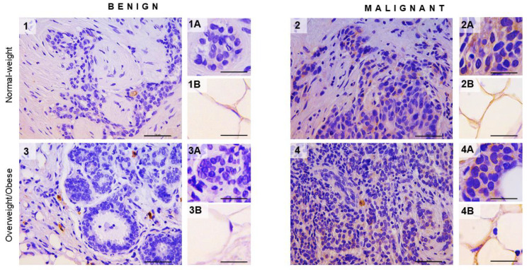
Figure 4.
Hexokinase 2 expression in breast tumor and associated adipose tissue in normal-weight and overweight/obesepremenopausal women. Immunohistochemistry and light microscopy showing hexokinase 2 protein localization patterns in benign (panels 1 and 3) and malignant (panels 2 and 4) tumor tissue. Cell-specific hexokinase 2 (brown staining) protein localization in breast epithelial cells (inserts 1A and 3A) and adipocytes (1B and 3B) in benign tumor tissues and breast cancer cells (inserts 2A and 4A) and cancer-associated adipocytes (2B and 4B) in malignant tumor tissues of normal-weight (panels 1 and 2) and overweight/obese (panels 3 and 4) premenopausal women. Magnification: ×40 and ×100, orig., respectively. Scale bars: 50 μm and 20 μm, respectively.

