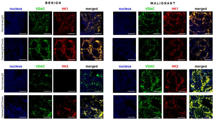
Figure 5.
Hexokinase 2 colocalization with mitochondrial marker VDAC in premenopausal breast cancer. Immunofluorescence staining and confocal microscopy images showing voltage-dependent anion-selective channel protein 1 (VDAC)/hexokinase 1 (HK 1) and VDAC/hexokinase 2 (HK 2) protein localization in benign and malignant breast tumor tissue of normal-weight and overweight/obese premenopausal women. Double immunolabeling is shown in separate channel micrographs against VDAC (green), HKs (red), and as merged channels showing colocalization of VDAC and HKs (yellow). Nuclei were stained with Sytox Orange (false blue). Magnification: ×63, Zoom: ×2. Scale bars: 10 μm.

