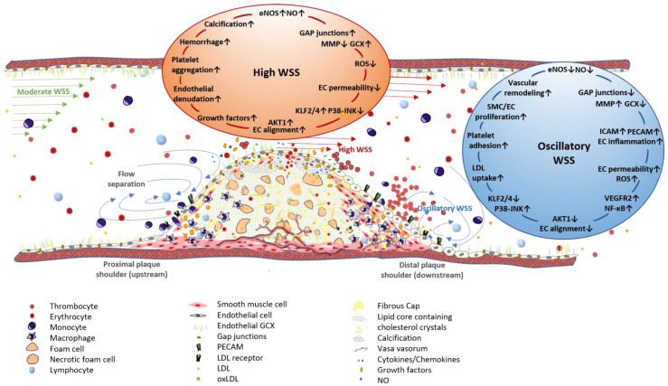
Figure 3.
Association of WSS patterns of the arterial wall with plaque progression and vulnerability. Vessel stenosis caused by an advanced atherosclerotic lesion changes local WSS patterns. Moderate WSS, to which straight vessel segments are exposed, changes to high WSS at the throat of an arterial obstruction, whereas the downstream plaque shoulder is exposed to oscillatory WSS, characterized by flow separation and bidirectional flow. The endothelium, affected by high WSS, exhibits enhanced NO production due to mechanoactivation of eNOS, which is induced by the antiinflammatory transcription factor KLF2. Additionally, ECs show an axial alignment in the direction of blood flow, an almost intact GLX, and the low expression levels of adhesion molecules, such as PECAM and ICAM-1. Apart from its potentially atheroprotective effects, high WSS can promote plaque vulnerability by inducing platelet aggregation and endothelial denudation. Furthermore, plaques in vessel segments exposed to high WSS are characterized by enhanced intimal calcification. Oscillatory WSS is involved in plaque initiation and progression through the induction of EC and SMC proliferation and migration. Mechanoactivation of NF-ᴋB results in the increased expression of diverse proinflammatory genes, i.e., adhesion molecules and receptors for LDL uptake. EC alignment is inhibited and cells show a cobblestone-like morphology and increased permeability. Enhanced eGCX shedding due to increased MMP or sheddase levels facilitates the infiltration of immune cells from peripheral blood into the vessel wall. EC: endothelial cell, eNOS: endothelial nitric oxide synthase, GCX: glycocalyx, ICAM-1: intercellular adhesion molecule 1, KLF2: Krüppel-like factor 2, LDL: low density lipoprotein, NO: nitic oxide, PECAM: platelet endothelial cell adhesion molecule, SMC: smooth muscle cell, WSS: wall shear stress, ↑: upregulation, ↓: downregulation.

