Figure 1.
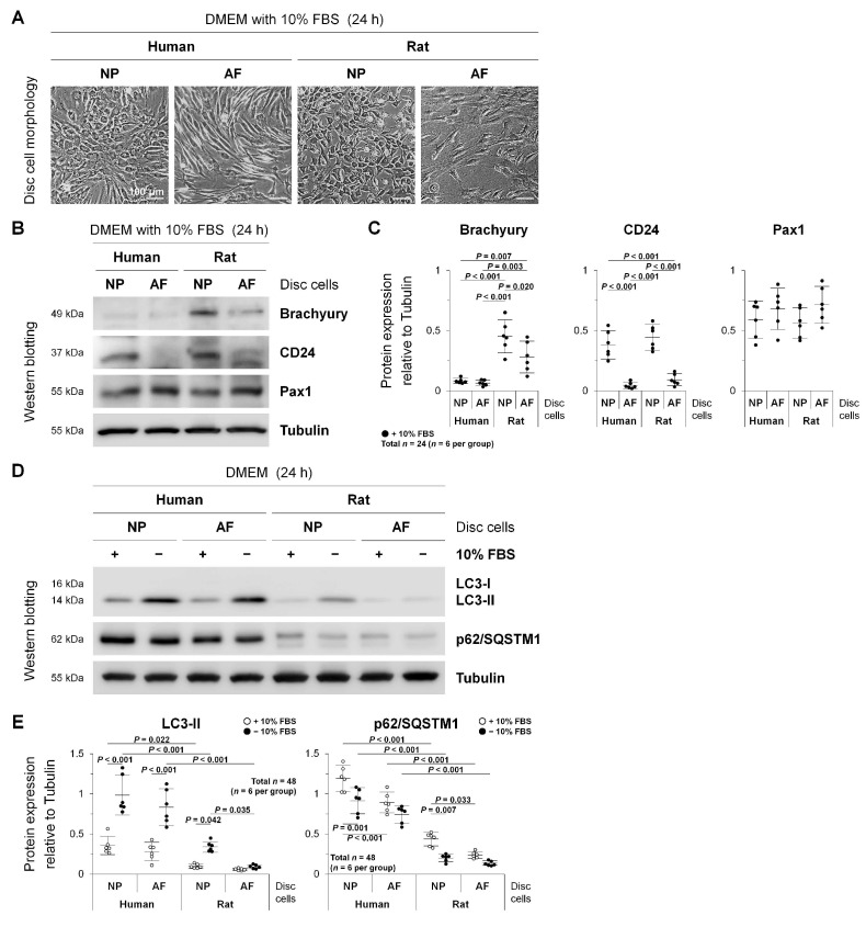
Autophagy in human degenerated and rat non-degenerated disc NP and AF cells. (A) Morphological appearance of human and rat lumbar disc NP and AF cells after 24 h culture in DMEM with 10% FBS. Cellular images (human NP and AF cells from the L4–L5 disc with the Pfirrmann grade 3 in a 64-year-old woman) are representative of experiments with similar results (n = 6). (B) Western blotting for NP notochordal markers brachyury and CD24 and NP and AF marker Pax1 in total protein extracts from human and rat lumbar disc NP and AF cells after 24 h culture in DMEM with 10% FBS. Tubulin was used as a loading control. Immunoblots shown (human NP and AF cells from the L4–L5 disc with the Pfirrmann grade 3 in a 64-year-old woman) are representative of experiments with similar results (n = 6). (C) Changes in protein expression of brachyury, CD24, and Pax1 relative to tubulin in human and rat lumbar disc NP and AF cell protein extracts. Data are the mean ± 95% CI. Two-way ANOVA with the Tukey–Kramer post-hoc test was used (n = 6). (D) Western blotting for autophagy marker LC3, autophagy substrate p62/SQSTM1, and loading control tubulin in total protein extracts from human and rat lumbar disc NP and AF cells after 24 h culture in DMEM with and without 10% FBS for serum deprivation. Immunoblots shown (human NP and AF cells from the L4–L5 disc with the Pfirrmann grade 3 in a 64-year-old woman) are representative of experiments with similar results (n = 6). (E) Changes in protein expression of LC3-II and p62/SQSTM1 relative to tubulin in human and rat lumbar disc NP and AF cell protein extracts. Data are the mean ± 95% CI. Two-way ANOVA with the Tukey–Kramer post-hoc test was used (n = 6).

