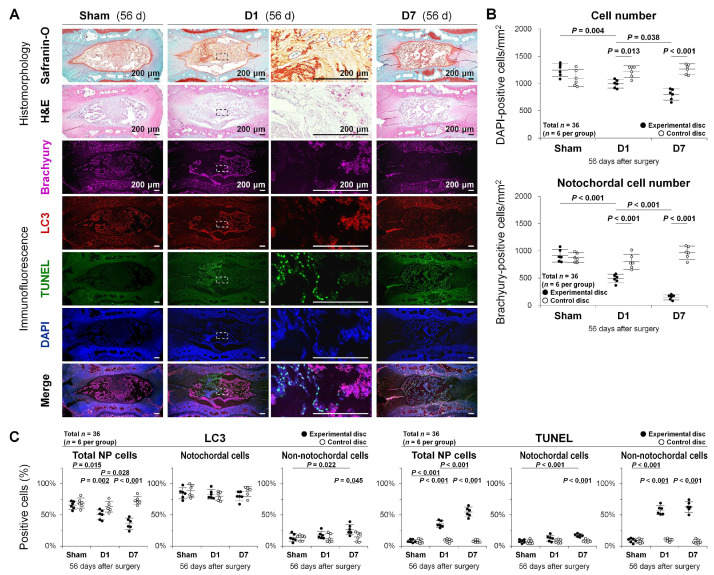
Figure 4.
Autophagy, apoptosis, and notochordal cell disappearance in rat tail temporary static compression-induced disc degeneration. (A) Safranin-O, fast green, and hematoxylin staining, H&E staining, and multi-color immunofluorescence for NP notochordal brachyury (purple), autophagic LC3 (red), apoptotic TUNEL (green), nuclear DAPI (blue), and merged signals in rat tail experimental disc NP sections at 56 days of 0-day (sham, unloaded throughout), 1-day (D1, loaded for the first 24 h and unloaded later), and 7-day (D7, loaded for the initial 7 days and unloaded later) temporary static compression. Immunoimages shown are representative of experiments with similar results (n = 6). (B) Changes in the number of DAPI-positive total cells and brachyury-positive notochordal cells in rat tail experimental and control disc NP sections at 56 days of 0-day (sham), 1-day (D1), and 7-day (D7) temporary static compression. Data are the mean ± 95% CI. Two-way ANOVA with the Tukey–Kramer post-hoc test was used (n = 6). (C) Changes in the percentage of LC3-positive cells and TUNEL-positive cells relative to DAPI-positive total cells, brachyury-positive notochordal cells, and brachyury-negative non-notochordal cells in rat tail experimental and control disc NP sections at 56 days of 0-day (sham), 1-day (D1), and 7-day (D7) temporary static compression. Data are the mean ± 95% CI. Two-way ANOVA with the Tukey–Kramer post-hoc test was used (n = 6).

