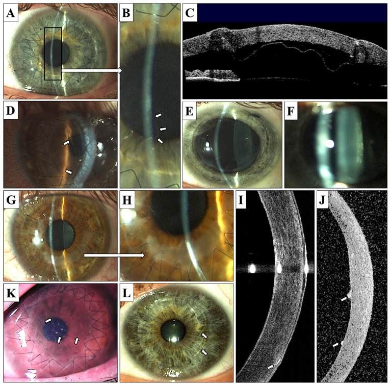
Figure 3.
Postoperative complications of DALK and PK surgery. (A–D)—DALK, Descemet’s membrane detachment; (A)—localized, mild detachment (magnification presented on photograph B, arrows), (C)—severe Descemet’s membrane detachment visualized in anterior OCT (Visante); (D)—severe Descemet’s membrane detachment causing corneal graft edema (arrows). (E)—PK, Urrets–Zavalia syndrome; (F)—PK, posterior subcapsular cataract formation; (G–I)—DALK, subepithelial opacifications (G,H)—opacifications visible in area of sutures, mostly in the lower cornea; (I)—anterior OCT Visante, subepithelial opacifications visible in lower cornea, arrow); (J)—PK, anterior OCT Visante, endothelial sediments associated with endothelial graft disease; (K)—PK, endothelial graft disease with visible Khodadoust line (arrows) and endothelial sediments; and (L)—PK, Descemet’s membrane folds (arrows).

