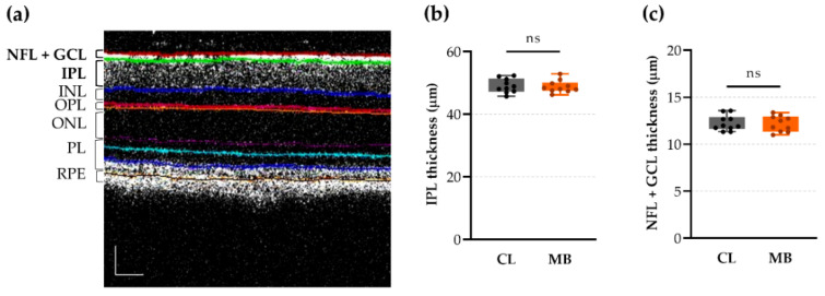
Figure 5.
In vivo quantification of retinal layer thickness via optical coherence tomography (OCT) revealed no changes at five weeks after microbead injection. (a) Representative OCT image with colored lines indicating the borders of different retinal layers. Scale bar = 50 µm. (b) The inner plexiform layer (IPL) thickness was unchanged upon microbead injection. Unpaired two-tailed t-test, ns = non-significant, n = 10. (c) Similarly, the thickness of the combined nerve fiber and ganglion cell layers (NFL + GCL) remained unaltered after microbead injection. Unpaired two-tailed t-test, ns = non-significant, n = 10. CL = contralateral eyes and MB = microbead-injected eyes.

