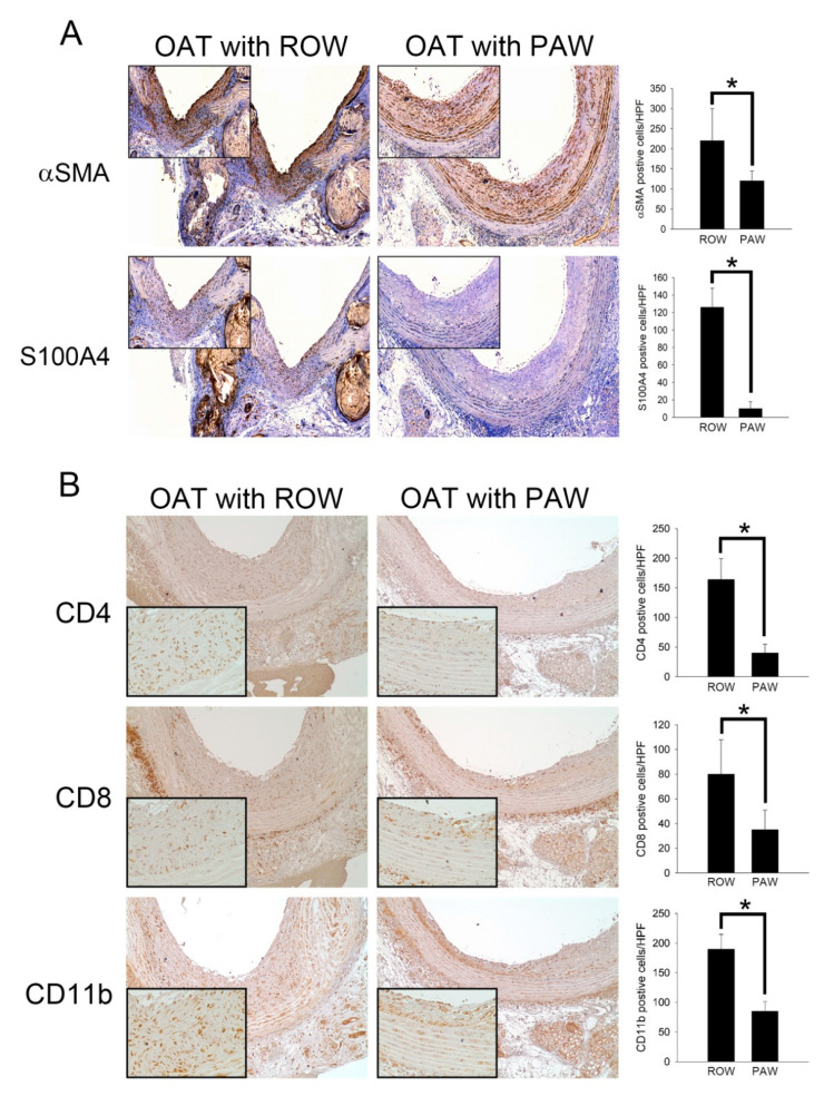
Figure 2.
Administration of PAW is effective against SMCs, fibroblasts, CD4+ T lymphocytes, CD8+ T lymphocytes, and macrophage activity in OAT-induced chronic allograft vasculopathy. (A) Immunohistochemistry to assess proliferated SMCs (αSMA) and accumulated fibroblasts (S100A4) in rat thoracic aortas from donor PVG/Seac rats. The lumen is uppermost in all sections; the images are 200× magnified. Similar regions are shown as enlarged images (400× magnification) in the upper left corners. The brown signal indicates αSMA- and S100A4-positivecells. (B) Immunohistochemistry to analyze accumulated helper T cells (CD4), cytotoxic T cells (CD8), and infiltrated macrophages (CD11b) in rat thoracic aortas from donor PVG/Seac rats. The images are 200× magnified. Similar regions are shown as enlarged images (400× magnification) in the lower left corners. The brown signal indicates CD4-, CD8- and CD11b-positive cells. Semi-quantification of immunohistochemically positive stained cells in high power field (HPF) is shown in the right panel of (A,B). The graphs represent the accumulation of cells in the aortas of rats from the experimental groups. The results are expressed as the mean ± SD. Statistical evaluations were performed using Student t-test, followed by Dunnett’s test. * p < 0.05 was considered as significant.

