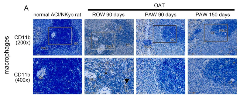
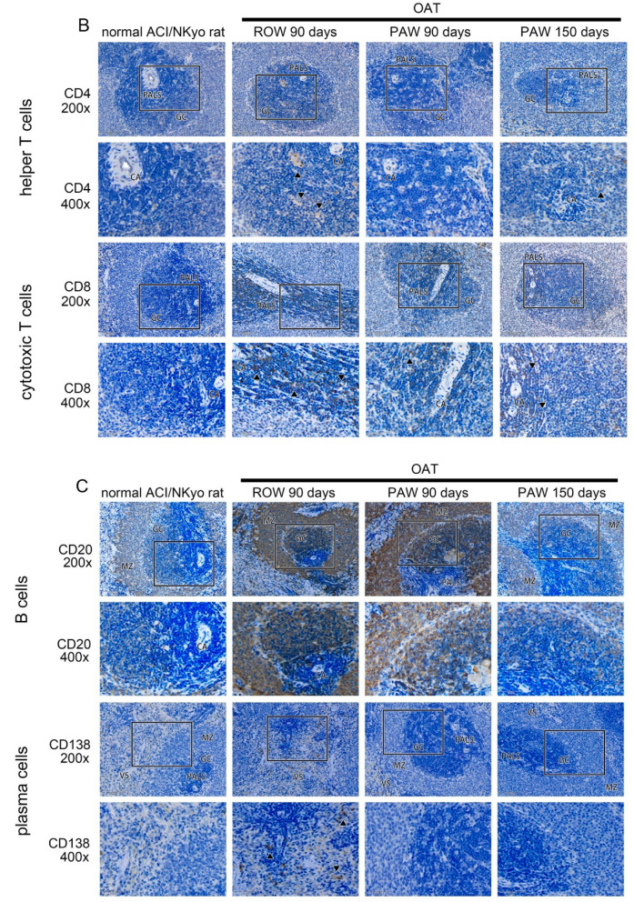
Figure 3.
Decreased activity of macrophages, T lymphocytes, B lymphocytes, and plasma cells in the spleen of PAW administered OAT-ACI/NKyo rats. (A) Immunohistochemistry was performed to assess the accumulated macrophages (CD11b) in the spleen of OAT-recipient ACI/NKyo rats (GC, germinal center; PALS, periarterial lymphatic sheath; CA, central artery). The brown signals indicated by triangle arrow heads are CD11b-positive macrophages. The images in the upper column are 200x magnified, and the square regions are shown as enlarged images (400× magnification) in the lower column. (B) Immunohistochemistry was performed to analyze accumulated helper T cells (CD4) and cytotoxic T cells (CD8) in the spleen of OAT-recipient ACI/NKyo rats. The images are 200× and 400× magnified, respectively. The brown signal indicated by triangle arrow heads areCD4- and CD8-positivecells. (C) Immunohistochemical analysis of accumulated B cells (CD20) and activated plasma cells (CD138) in the spleen of OAT-recipient ACI/NKyo rats (MZ, mantle zone; VS, venous sinuses). The images are 200× and 400× magnified, respectively. The brown signals indicated by triangle arrow heads are CD138-positivecells. The cell nuclei were stained with hematoxylin, and the slides were observed via light microscopy.


