Figure 5.
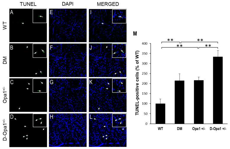
Reduced Opa1 level exacerbates diabetes-induced apoptosis of vascular cells in retinal capillary networks. Representative images show TUNEL-positive cells (arrows) in retinal capillaries of (A) WT mice, (B) diabetic (DM) mice, (C) Opa1+/− mice, and (D) diabetic Opa1+/− mice (D-Opa1+/−). (E–H) Corresponding images of DAPI-stained cells in the retinal capillary networks, respectively. (I–L) Merged images show TUNEL-positive cells are superimposed with DAPI-stained cells; 20× magnification. Insets represent enlarged images of TUNEL-positive cells. (M) Graph representing cumulative data shows increased number of TUNEL-positive cells in retinal capillaries of diabetic mice compared with that of WT mice, while retinal capillary networks of D-Opa1+/− mice show a further increase in the number of TUNEL-positive cells compared with that of diabetic mice. Data are presented as mean ± SD. ** = p < 0.05, n = 6.

